From the pages you shared (first chapter of Anatomy of Eye in Comprehensive Ophthalmology – A.K. Khurana), examiners usually ask “named concepts” rather than long paragraphs. These are short definitions or anatomical terms that carry a specific name.
Below are the high-yield named concepts from these pages that MBBS examiners frequently ask in viva/short notes.
Important Named Concepts (Anatomy of Eye)
1. Poles of the Eyeball


Definition:
The poles are the two central points of the maximal curvature of the eyeball.
Types
Anterior pole – centre of cornea
Posterior pole – centre of posterior curvature of sclera
Related concept
Axis of eyeball – imaginary line joining anterior and posterior poles.
Examiner favourite viva question:
"What are the poles of the eyeball?"
2. Equator of the Eyeball
Definition:
The equator is an imaginary circular line midway between the anterior and posterior poles of the eyeball.
Importance
Divides eyeball into anterior half and posterior half.
Used as a reference point in ophthalmic surgery and anatomy.
3. Ora Serrata

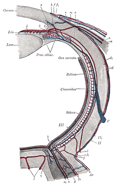
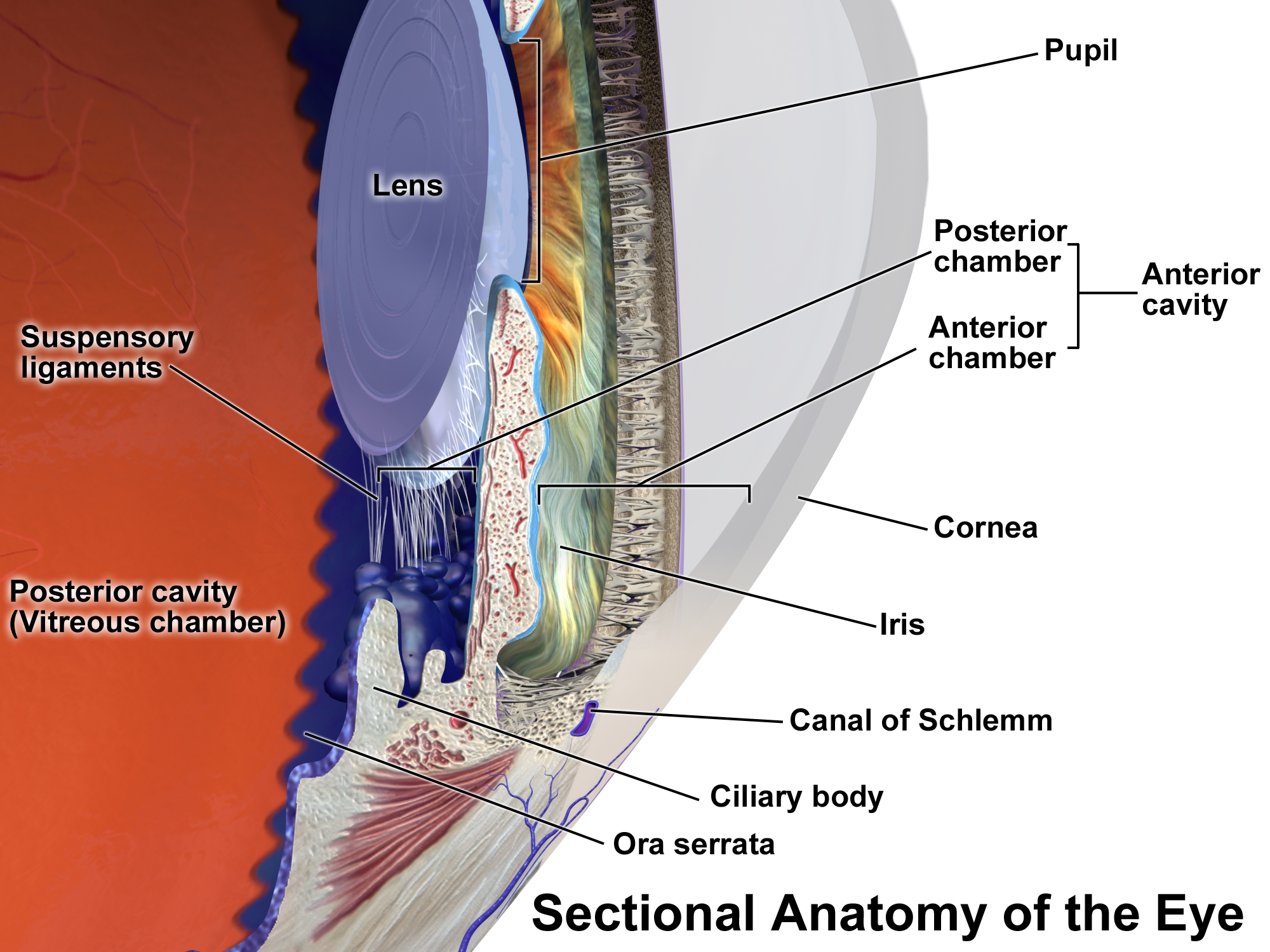

Definition:
The ora serrata is the anterior serrated margin of the retina where the retina ends and the ciliary body begins.
Key points
Marks the transition from photosensitive retina → non-photosensitive epithelium
Appears toothed or serrated
Exam question:
"What is ora serrata?"
4. Limbus


Definition:
The limbus is the junction between the cornea and sclera.
Important facts
Conjunctiva firmly attaches here
Important surgical landmark
Contains limbal stem cells
Examiner favourite:
"Define limbus."
5. Coats of the Eyeball


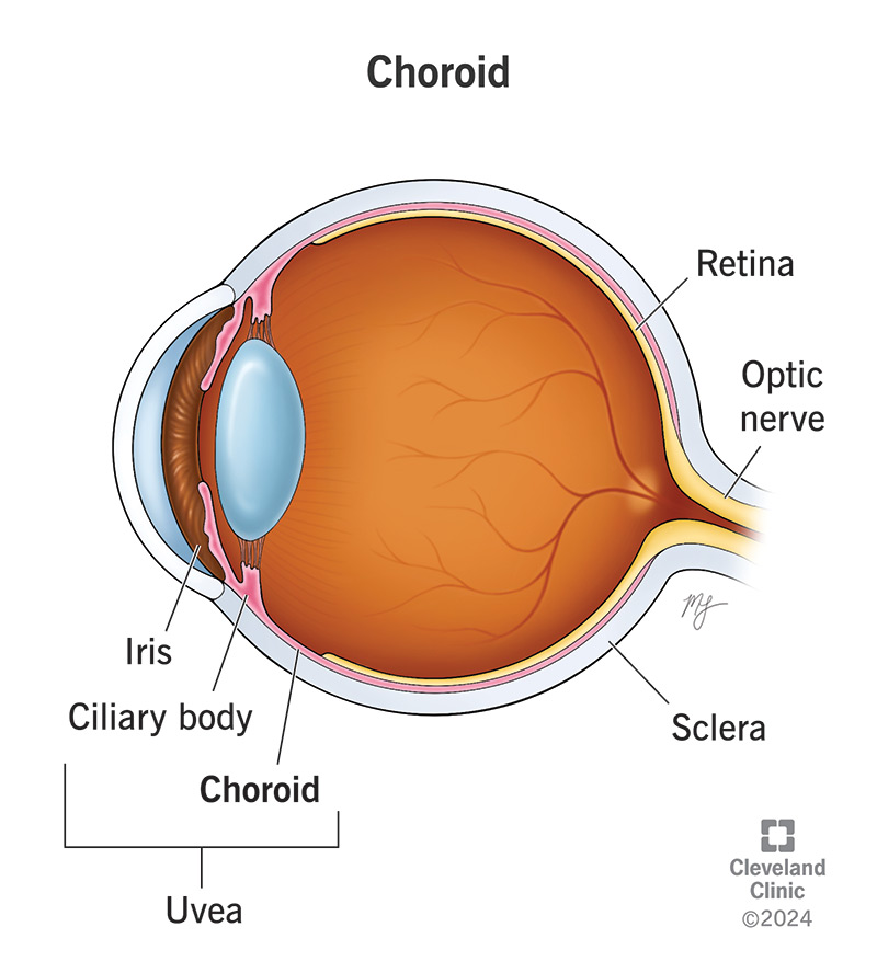
Eyeball has 3 coats (tunics).
1. Fibrous coat
Cornea
Sclera
Function: protection and shape.
2. Vascular coat (Uveal tract)
Iris
Ciliary body
Choroid
Function: nutrition and accommodation.
3. Nervous coat
Retina
Function: vision.
Exam question:
"Name the coats of the eyeball."
6. Anterior Chamber
Definition:
The space between cornea and iris filled with aqueous humour.
Boundaries
Anterior → posterior surface of cornea
Posterior → anterior surface of iris
Normal depth
About 2.5 mm
Volume of aqueous humour
~ 0.25 ml
7. Posterior Chamber
Definition:
The triangular space between iris and lens.
Boundaries
Anterior → posterior surface of iris
Posterior → lens and zonules
Lateral → ciliary body
Contains aqueous humour.
8. Segments of the Eyeball
The eye is divided into two segments.
1. Anterior segment
Contains:
Cornea
Iris
Lens
Aqueous humour
Anterior chamber
Posterior chamber
2. Posterior segment
Contains:
Vitreous humour
Retina
Choroid
Optic disc
9. Appendages of the Eye
Structures that protect and support the eyeball.
Includes:
Eyelids
Eyebrows
Conjunctiva
Lacrimal apparatus
Exam question:
"What are appendages of the eye?"
10. Visual Pathway


Definition:
The pathway through which visual impulses travel from retina to the brain.
Sequence
Retina
Optic nerve
Optic chiasma
Optic tract
Lateral geniculate body
Optic radiation
Visual cortex (occipital lobe)
Ultra-High Yield Viva One-liners
Memorize these exactly.
Shape of eyeball: Oblate spheroid
AP diameter: 24 mm
Horizontal diameter: 23.5 mm
Vertical diameter: 23 mm
Volume: 6.5 ml
Weight: ~7 g
✅ Most asked in MBBS viva from this chapter
Poles of eyeball
Equator of eyeball
Ora serrata
Limbus
Coats of eyeball
Anterior vs posterior chamber
Segments of eyeball
Visual pathway
If you want, I can also give the “Top 20 examiner favourite named concepts from the whole Ophthalmology book” that come repeatedly in MBBS viva and NEET-PG.
Comments
Post a Comment